

Differential Technologies
Fisaude Occasion
Best Deals
Our Kinefis products
News
Offers
Outlet
Fisaude Tech Academy
Physiotherapy
Chiropody
Aesthetics, dermocosmetics and aesthetic medicine
Wellness, quality of life and body care
Odontology
Medical equipment
Chinese traditional medicine
Clinical furniture
Therapeutic cabinets
Essential protection material for coronaviruses
Aerobics, fitness and pilates
Sports and games
Sanitary wardrobes
Veterinary
Orthopedics
Surgical instruments (clearance)
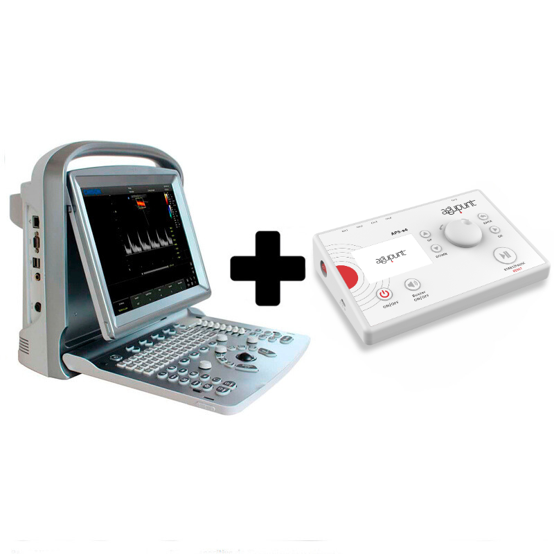
Savings pack: Chison ECO 6 ultrasound machine with 10 MHz linear probe + APS e4 therapeutic percutaneous electrolysis device

Check delivery time
Description
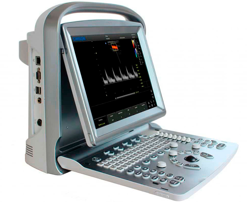
Chison ECO 6 ultrasound machine with 10 MHz linear probe + APS e4 therapeutic percutaneous electrolysis device
Chison ECO 6 portable ultrasound machine
- The most powerful device with the best image quality in the ECO family
- Integrated battery lasting up to 3 hours for easy mobility
- Multitude of clinical applications and powerful PW
- New smart control panel: more comfortable, functional and intuitive
- Innovative and functional needle software
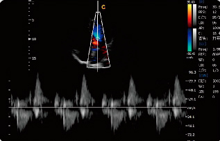
- Color Doppler (different color units, depending on the speed and direction of blood flow)
- Power Doppler (displays color information about the amplitude of the Doppler signal - non-directional)
The family of Chison ECO ultrasound machines adds a new member. A device that multiplies the virtues of these ultrasound machines to become the most powerful device, with more possibilities of action (it has a large number of clinical applications) and with the best image quality of the entire ECO range. And that is a lot to say.
The ECO 1 has made a name for itself as one of the best-selling ultrasound machines in the mid-range. The ECO 2 has marked a before and after in the field of health thanks to its premium features at a mid-range price. The ECO 5 has become one of the best options for a color Doppler ultrasound system with advanced technology at a competitive price...and now the ECO 6 arrives on the European market with all the virtues of its predecessors and with a large arsenal of new resources.
As with the rest of the ECO ultrasound machines, the ECO 6 model manages to stand out from the rest of the ultrasound machines on the market since it combines the best of two worlds: It offers state-of-the-art features that adapt like a glove to the work of doctors and physiotherapists and does so at a much more than competitive price.
Thus, its intelligent control panel design, the full screen option, the ergonomic LED screen, the patented EasyViewTM technology, the backlit keyboard, the magnificent PW resolution, join forces to transform the ECO 2 into an unattainable device for the rest of the mid-range ultrasound machines. The reference device. An exceptional and incomparable ultrasound machine that has come to reign in the world of mid-range ultrasound machines. He has plenty of virtues.
ECO 6 Ultrasound Technologies:
1) FHI: Fusion Harmonic Imaging , maximizes image quality and resolution without losing penetration, a technology that is especially positive when performing deep scans of the patient.
2) AIO: Automatic Image Optimization , in B mode, is used to optimize the distorted 2D image to preset the gain value; In PW or CW mode, it is used to optimize the spectrum baseline to the preset position.
3) Q-image: A series of innovative algorithms manage to improve image quality threefold. In addition, the set of chips that make up the ultrasound machine ensure pixel speed.
4) SRA & Compound Image: Highly flexible and cost-effective imaging tool that allows operators to observe and record images of a wide variety of anatomical sections in real time.
5) Q-beam: Improves image processing speed by improving signal transmission and receiving speed.
6) Chroma: Greatly improves contrast resolution by improving signal-to-noise ratio.
Main Functions of ECO 6:
- Real-time automatic tracing: In PW mode, it can trace the spectrum in real time and calculate RI, PI and HR automatically. Traditional tracing is done after freezing, while automatic tracing will show all data in real time, without the need for freezing. Save time and improve work efficiency and accurate calculation results.
- TDI: Tissue Doppler Imaging , One of the functions that brings out the most performance from the Chison ECO 6 ultrasound machine. The TDI function is used to detect the movement of the myocardium and to analyze the systolic and diastolic function of the human body. For example, if there is a false positive diastolic function, the TDI level decreases.
- Color M Mode: Doppler mode within the M mode image, provides color-coded qualitative information of fluid movement. Shows cardiac motion and blood flow information at the same time. If there is mitral regurgitation, we use Color M mode to see the direction of the blood when the mitral valve closes.
- 2D Steer: It is based on huyghens principle, directs the ultrasound beam to enlarge the image area in the far field, and can also focus the ultrasound beam to improve the axial resolution, which is good for scanning of nerve, tendon and small deep vessels. 2D Steer deflects the ultrasound beam by 20 degrees to receive more information from the reflected echo and improve the visualization of tissue information.
- B/BC Mode: Shows 2 images at the same time. One is in B mode and the other is in C mode. So you can get the B mode information and blood flow information at the same time. For example, if there is a carotid plague, the doctor can measure the distance of the plague and see the blood information at the same time. 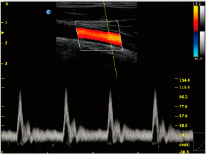
- Triplex: Shows B, C and PW mode together in real time. Let the doctor find the target tissue and display the spectrum very easily, without needing to go back to B mode or C mode and find the target tissue again. For example, when the doctor scans the carotid artery, if the image is zoomed out, the doctor can move the sample gate to obtain the spectrum directly.
- Supper Needle: Uses an acoustic beam fusion method based on the normal image to change the direction of the ultrasound beam and uses a low mechanical index to improve metal visualization. Super Needle helps improve needle visualization during procedures. The needle line is adjustable by 30 degrees on each side, better for getting closer to the deep container.
- CW: Continuous wave Doppler, used to measure blood at high speed in the heart. If it is mitral valve regurgitation, CW mode can measure the velocity and differentiate between philological regurgitation and pathological regurgitation.
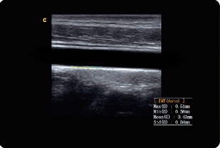
- Auto IMT: The IMT is the internal thickness of muscles and joints. With this function, the doctor can measure the IMT automatically and obtain three results at the same time. A function that makes this type of measurement faster, easier and more precise. A function that is especially beneficial for medical work since the thickness of the carotid is very important for atherosclerosis to be diagnosed.
- Trapeze: only used in linear probe. It can help increase the scanning range.
- CPA & DPD.
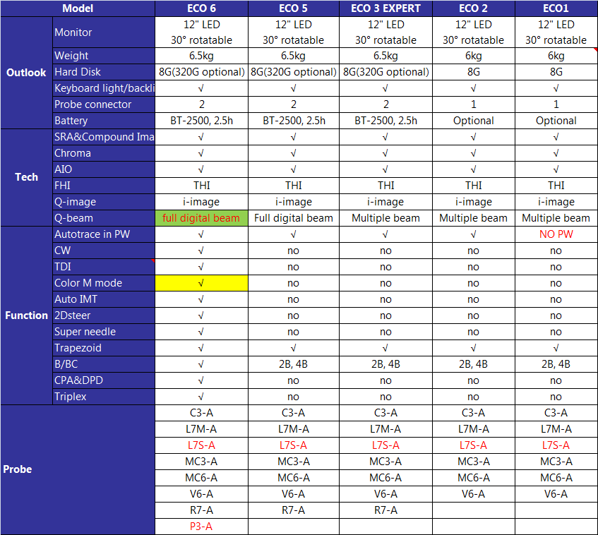
Comparative table of Chison ultrasound machines:
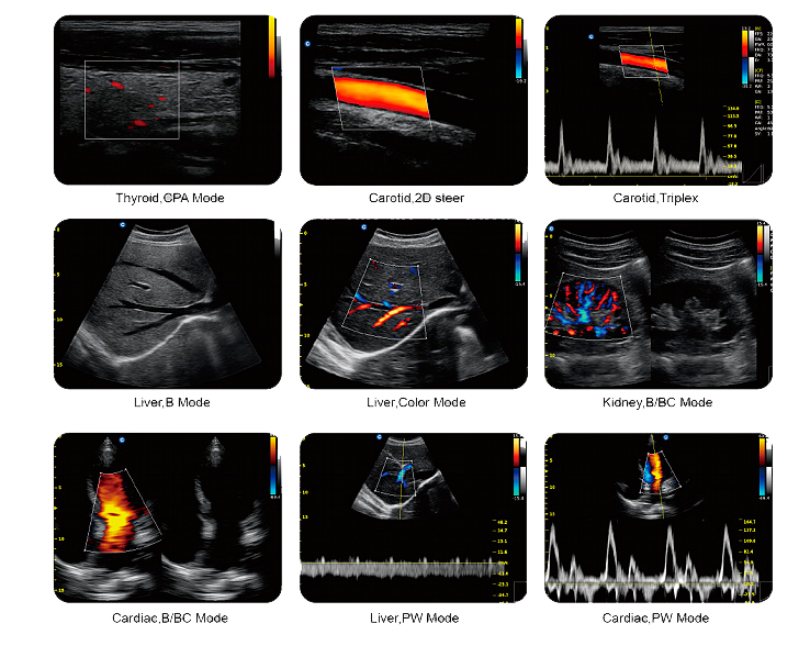
Graphic map of the Chison ECO 6 functions:
Technical characteristics:
- Main applications: Abdomen, OB / GYN, Vascular, small and specific areas (breasts, testicles, thyroid), conventional and superficial musculoskeletal, pediatric, basic cardiac
- Imaging technologies: i-Image, SRA, Compound, THI
- Display modes: B, B/B, 4B, M, B/M, PW
- Grayscale: Color M Mode
- Monitor: 12 inches - 30º rotation
- Scanning depth: 240 mm max.
- Probe frequency: 10MHz
- Zoom: 4 steps
- Software Package: General, OB/GYN, URO, Cardiac, Vascular, Small Parts
- File management: patient information management, image registration, examination report
- Power supply: AC 100V-240V, 50Hz-60Hz
- Full Screen Mode: i-Image, Chroma, SRA, Composite Images, THI,
- Memory card: 8G, PW mode
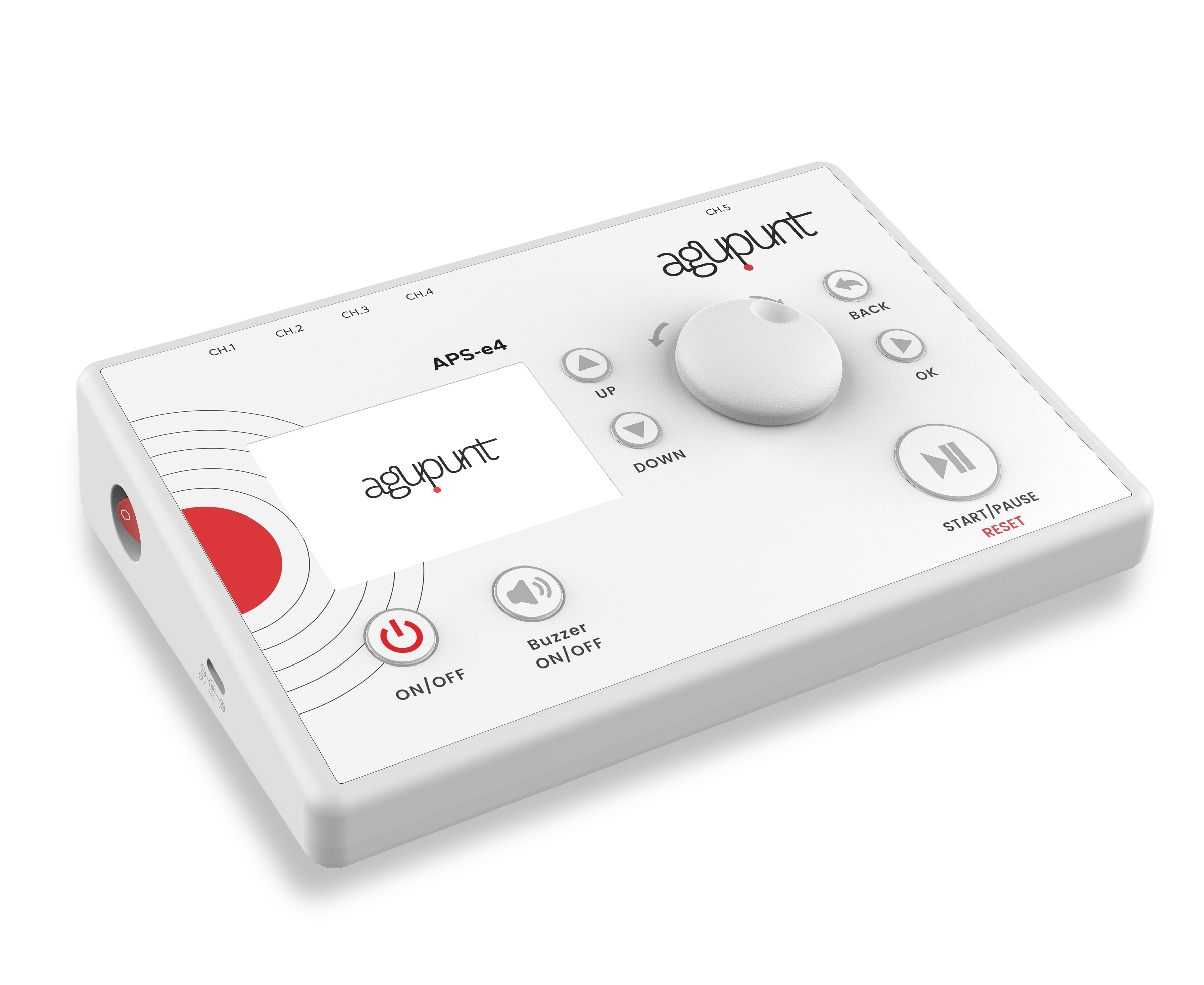
APS e4 Therapeutic percutaneous electrolysis: The revolution in invasive physiotherapy
The new APS e4 device has been redesigned and improved by the best professionals in invasive electrotherapy treatments. Includes the new 8-output octopus-type cable for electrolysis . A portable multi-option electrotherapy device with three types of current: galvanic (continuous), biphasic (symmetrical/asymmetrical) and monophasic (symmetrical). Its treatments are 100% modifiable, and allows you to control the parameters of all treatments, and adapt to the needs of each patient.
This device is based on the most innovative technology to redefine the field of invasive physical therapy. The APS e4 device erases the technological boundaries that limit other devices to elevate invasive physiotherapy to a new level.
Its technological components and the precision of its treatments combine to configure the most complete equipment on the market thanks, also, to its four work programs:
- Single-phase pulsed current (EPS, PENS)
- Percutaneous electrolysis (IPEN)
- Neuromodulation (NMP)
- Segmental stimulation (MSUN)
It is the only invasive physiotherapy equipment licensed to work on battery or connected to the electrical grid.
*Compatible with the previous model of APS-e4 and other electrolysis equipment.
Current typology
- Galvanic (continuous)
- Bisaphic (symmatric)
- Bisaphic (asymmetrical)
- Single phase (symmetrical)
Type of treatments
Parameters 100% modifiable during treatment to adapt them to each patient
- Dry electropuncture: Application to myofascial trigger points. Default programs and
Modifiable high and low frequency.
- Neuromodulation: Stimulation of dermatomes, myotomes and/or sclerotomas related to the pain area. Default and modifiable program.
- TENS: Analgesia induced by neurostimulation for the relief or blocking of chronic or acute pain.
- Microcurrents: Application at a subsensory level with multiple benefits for people with hypersensitivity.
- Pointer: Checking and stimulating the target tissue using a single needle.
- IPEN (electrolysis): Indicated for tendinopathies and muscle tissue. It produces electrochemical changes in the tissue that stimulate regenerative responses.
Strengths:
- INCLUDES 8-HOUR IN-PERSON TRAINING. CHECK THE DATES OF THE NEXT COURSES BY CLICKING HERE.
- The equipment has 4 types of currents: 1. Pulsed monophasic current (EPS, PENS), 2. Percutaneous electrolysis (IPEN), 3. Neuromodulation (NMP), 4. Segmental stimulation (MSUN).
- Never has a laptop with these characteristics taken up so little space.
- APS e4 offers maximum stability and precision in the emission of currents. In addition, it allows us to know the exact voltage load that we are emitting in order to assess the real effectiveness of the treatments.
- It is the first invasive physiotherapy equipment with which you can work connected to the electrical network. With a health license to be able to work plugged into the electrical network.
- The equipment has an anti-collapse system that automatically restarts it in case of blocking, thus avoiding interfering with the patient.
- It has 50 storage memories to create your treatments.
- APS e4 has a pointer capable of emitting current superficially using a strut and 4 channels with crocodile clips to be able to emit currents percutaneously with a needle.
- The checkbox system performs a constant self-check of the equipment and its connections.
- The variety of programs included in the device allow for a multitude of treatments.
What is invasive physiotherapy?
Invasive physiotherapy is a percutaneous intratissue electrolysis (EPI) technique that consists of the ultrasound-guided percutaneous application of a galvanic current through an acupuncture needle. This achieves phagocytosis and the repair of soft tissue injuries (patellar tendon, Achilles tendon, pubalgia/hernia, lateral and medial epicondylitis, etc.).
Who is this product aimed at?
To physiotherapy and rehabilitation clinics.
For what treatments is invasive physiotherapy recommended?
- Tendinopathies: such as in the supraspinatus, rotator cuff, epicondylitis, patellar tendinopathy, Achilles tendinopathy, etc...
- Muscle tears
- Neuropathies such as loss of sensitivity or motor deficit, etc...
Technical characteristics:
- Rechargeable battery with USB type C charger (universal)
- Operation of the equipment connected to the current or by battery.
- Outputs 1-4 of symmetrical monophasic, symmetrical biphasic and asymmetrical biphasic current for dry electropuncture, neuromodulation, TENS and microcurrent treatments.
- Output 5 for galvanic current (for electrolysis) and symmetrical two-phase (for pointer).
- 3.5 inch TFT digital screen.
- Easy and comfortable to carry, with dimensions of 220m x 140 mm and a weight of 225g.
- Acoustic sounds (start and end of treatment or in case of equipment error)
- Possibility of working in milliamps (mA) or microamps (A) in the electrolysis and microcurrent programs.
- Equipment parameters:
Power: 0.01 to 20.0 mA
Frequency: 1 to 150 Hz
Pulse: 10 to 700 ms
Stage time: from 1 to 60 sec
Treatment time: 1 sec to 60 min
Maximum voltage: 19V
Dimensions:
- Weight: 2 kg.
Base equipment:
- Carrying case with APSe4
- Handle with on and off button
- 4 cables for channels 1-4
- Charger cable designed for medical device
- Pointer for the maniple
- 4 electrodes
- 25 APS Regular needles 0.32x40 mm
- Octopus type cable with 8 outputs
Warranty:
- The device has a two-year warranty.


