

Differential Technologies
Fisaude Occasion
Best Deals
Our Kinefis products
News
Offers
Outlet
Fisaude Tech Academy
Physiotherapy
Chiropody
Aesthetics, dermocosmetics and aesthetic medicine
Wellness, quality of life and body care
Odontology
Medical equipment
Chinese traditional medicine
Clinical furniture
Therapeutic cabinets
Essential protection material for coronaviruses
Aerobics, fitness and pilates
Sports and games
Sanitary wardrobes
Veterinary
Orthopedics
Surgical instruments (clearance)
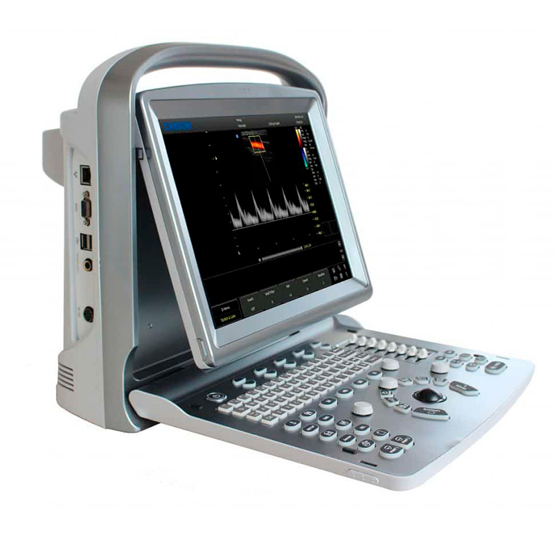
Chison ECO6 Portable Ultrasound Machine

Shipping in 4-5 weeks approx.
Description
Chison ECO 6 portable ultrasound machine
- The most powerful device with the best image quality in the ECO family
- Built-in battery life of up to 3 hours for easy mobility
- Multitude of clinical applications and powerful PW
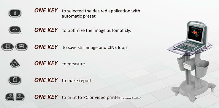
- New smart control panel: more comfortable, functional and intuitive
The Chison ECO ultrasound family has a new member. A device that multiplies the virtues of these ultrasound machines to establish itself as the most powerful device, with more possibilities of action (it has a large number of clinical applications) and with better image quality . the entire ECO range. And that's saying a lot.
The ECO 1 has made a name for itself as one of the best-selling ultrasound machines in the mid-range. The ECO 2 has marked a before and after in the health field thanks to its premium features at a mid-range price. The ECO 5 It has become one of the best options for color Doppler ultrasound systems with advanced technology at a competitive price...and now the ECO 6 reaches the European market with all the virtues of its predecessors and with a large arsenal of new resources.
As with the rest of the ECO ultrasound scanners, the ECO 6 model stands out from the rest of the ultrasound scanners on the market because it combines the best of both worlds: It offers cutting-edge features that fit like a glove to the work of doctors and physiotherapists and does so at a much more than competitive price.
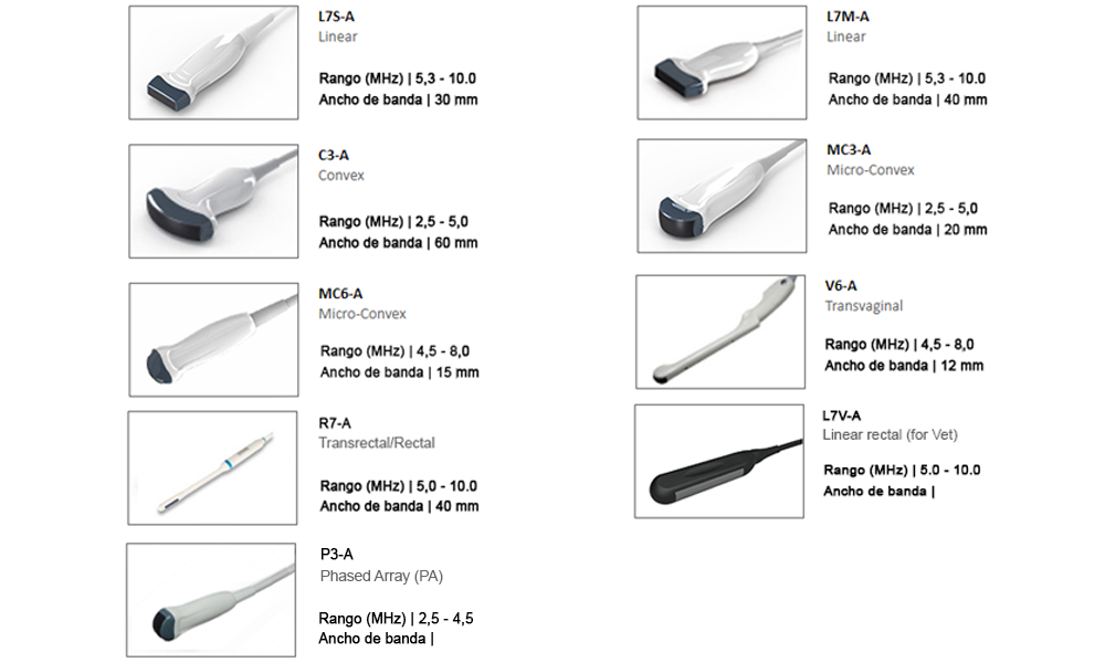
Choose the probe to equip your ECO 6 ultrasound scanner
Chison ECO 6 ultrasound technologies:
1) FHI: Fusion Harmonic Imaging , maximizes image quality and resolution without losing penetration, a technology that is especially positive when performing deep scans of the patient.
2) AIO: Automatic Image Optimization , in B mode, it is used to optimize the distorted 2D image to preset gain value; in PW or CW mode, it is used to optimize the spectrum baseline to preset position.
3) Q-image: A series of innovative algorithms improve image quality threefold. In addition, the set of chips that make up the ultrasound machine ensures pixel speed.
4) Q-flow: Maximizes the color sensitivity of low velocity blood flow or deep vessels.
5) Q-beam: Improves image processing speed by improving signal transmission and receiving speed.
6) X-contrast: Greatly improves contrast resolution by improving the signal-to-noise ratio.
Main Functions of ECO 6:
1) CW: Continuous wave Doppler, used to measure high velocity blood in the heart. If it is mitral valve regurgitation, CW mode can measure the velocity and differentiate between phylologic regurgitation and pathological regurgitation.
2) HPRF: High Pulse Repetition Frequency and has two sample gates for precise positioning of high velocity blood flow , similar to CW function. But the velocity it can detect is not as high as CW.
3) TDI: Tissue Doppler Imaging, is used to detect myocardial motion and to analyze left and right systolic and diastolic function. For example, if there is a false positive diastolic function decreases.
4) Color M Mode: Doppler mode within M mode imaging, provides color-coded qualitative information of fluid motion. It displays heart motion and blood flow information at the same time. If there is mitral regurgitation, we use Color M mode to see the direction of blood when the mitral valve closes.
5) ECG: A 3-lead ECG can be connected to display the real-time ECG wave of patients on the screen, providing more diagnostic information for the doctor.
6) Free direction M mode: Provides 3 sample lines on the screen at the same time. Each line can rotate 360 degrees. A very simple function that saves a lot of time for doctors.
7) Stress Echo: Uses ultrasound images of the heart to assess wall motion in response to physical stress. It helps to confirm or rule out the presence of coronary artery disease. Because patients with coronary artery blockages may have few or no symptoms at rest. Symptoms and signs of heart disease may be unmasked by exposing the heart to excise stress. In stress echo, we offer default templates and user-defined templates including quad mode real time recording and reporting.
8) Elastography: Displays tissue stiffness in real time to provide additional diagnostic information when scanning organs such as the breast. It is primarily used to display tumor elasticity as a replacement for physician palpation. Strain ratio measurement quantitatively provides the relationship between the average strain of the selected region and the nearby normal tissue region.
9) 18MHz Linear Probe: The maximum frequency of the probe can be up to 18MHz. It is designed for surface examination, providing superior detail resolution and contrast resolution , while the penetration can be up to 8cm.
10) Super Needle: It uses acoustic beam fusion method based on normal imaging, to change the direction of ultrasound beam and uses low mechanical index to improve visualization of metal. Super Needle helps to improve needle visualization during procedures. The needle line is adjustable in 30 degrees on each side, better to approach deep vessel.
11) 2D Steer: It is based on huyghens principle, it steers the ultrasound beam to enlarge the imaging area in far field, and also can focus the ultrasound beam to improve the axial resolution, which is good for scanning nerve, tendon and deep small vessels. 2D Steer deflects the ultrasonic beam by 20 degrees, so as to receive more information from reflected echo and improve the tissue visualization information. 2D steering can not only improve the image quality such as small vessels, nerves and MSK, but also can improve the visualization of needle. However, due to the low contrast, the needle enhancement is not as good as super needle.
12) Real-time auto-trace: In PW mode, it can trace the spectrum in real time and calculate RI, PI and HR automatically. Traditional trace is done after freezing, while auto-trace will display all data in real time, no need to freeze. It saves time and improves work efficiency and accurate calculation results.
13) B/BC Mode: It displays 2 images at the same time. One is in B mode and the other is in C mode. So you can get the B mode information and blood flow information at the same time. For example, if there is a carotid plague, the doctor can measure the plague distance and see the blood information at the same time.
14) Triplex: Display B, C and PW mode together in real time. Let doctor find the target tissue and display the spectrum very easily, no need to switch back to B mode or C mode and find the target tissue again. For example, when doctor scans carotid artery, if the image is zoomed out, doctor can move the sample gate to get the spectrum directly.
15) Duadplex: Combines B, C and PW mode with automatic tracking and measurement together in real time to help doctor make diagnosis more conveniently and accurately. For example, you can measure the umbilical artery of fetus in quadriplex mode.
16) Real-time panoramic imaging: It uses pattern recognition and image synthesis to generate and flexible viewing to reveal more anatomical information for diagnosis. It can only be used in linear probe, and you can expand the scanning width in real-time scanning as long as you want. For example, it is very useful when doctor tries to show the whole thyroid in one image.
17) Auto IMT: IMT stands for Intimate Thickness. The device can measure IMT automatically, with three results at the same time. This makes intimacy measurement faster, easier and more accurate.
18) Trapezoid: Only used on linear probe. It can help to increase the scanning range.
19) Automatic Follicle Tracking: Automatically detects follicles and provides efficient follicle sizes for IVF procedure.
20) Multi slice: Cut the 3D volume into different sectors, specialized for deep diagnosis .
Technical characteristics:
- Main applications: Abdomen, OB/GYN, Vascular, small and specific areas (breasts, testicles, thyroid), conventional and superficial musculoskeletal, pediatric, basic cardiac
- Imaging technologies: i-Image, SRA, Compound, THI
- Display modes: B, B/B, 4B, M, B/M, PW
- Grayscale: Color Mode M
- Monitor: 12 inches - Rotation 30º
- Scanning depth: 240 mm max.
- Probe frequency: 10MHz
- Zoom: 4 steps
- Software Package: General, OB/GYN, URO, Cardiac, Vascular, Small Parts
- File management: patient information management, image record, examination report
- Power supply: AC 100V-240V, 50Hz-60Hz
- Full screen mode: i-Image, Chroma, SRA, Composite images, THI,
- Memory card: 8G, PW mode
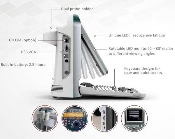
New design, more comfortable, functional and intuitive
Standard configuration:
- Chison ECO 6 device
- 3 USB ports
- 1 x Probe connector
- Includes 10MHz linear probe.
- Optional battery included.
- VGA port.
- Video port.
- LAN port.
Languages:
- Spanish, Italian, English, German, French, Chinese, Russian, Turkish and Czech.
Warranty:
The device has a 1 year warranty.
Reviews
4,6
")
")
8 reviews
 loading reviews...
Misael C.
Spain
19/12/2023

Diego M.
Spain
13/12/2023
Bien
Alba G.
Spain
04/12/2023
Me gusta
Susana M.
Spain
26/11/2023
Bueno
João G.
Spain
17/10/2023
Bons
Joaquim G.
Spain
15/10/2023
Muitos simples de trabalhar
Diego M.
Spain
24/09/2023
Muy completo y práctico
anonymous
Spain
12/01/2018
Un ecografo potente, manejable, de uso muy intuitivo


Me gusta, buena calidad